Dundee study finds new imaging method could help breast cancer patients avoid extensive surgery
Published On Wed 4 Dec 2019 by Dominic Glasgow
- University of Dundee scientists find new technique accurately identifies whether breast cancer cells remain after chemotherapy, and could help patients avoid more extensive surgery
- New ultrasound imaging method found to be as accurate as MRI in assessing the effectiveness of pre-surgery chemotherapy for breast cancer patients – and could be faster and cheaper for NHS
A new type of ultrasound imaging to assess the effectiveness of chemotherapy could help some breast cancer patients avoid more extensive surgery, a Scottish study has found.
The study, led by researchers from the University's School of Medicine, has found that combining shear wave elastography (SWE) with standard ultrasound (US) accurately measured how cancers respond to chemotherapy given before surgery (neo-adjuvant chemotherapy) and could help reduce the extent of patients’ surgery.
It is also hoped that shear wave elastography with ultrasound could prove a faster, cheaper and more widely-accessible method for the NHS compared to MRI, with results in less than five minutes.
Breast cancer is the most common cancer in the UK, with around 4,700 women in Scotland being diagnosed and over 1,000 people in Scotland still losing their lives to the disease each year.
Chemotherapy can be given before surgery to try to shrink the tumour in the breast, which in some cases can allow the patient to have breast-conserving surgery rather than a mastectomy. Chemotherapy before surgery could also be given to patients with rapidly growing tumours to help slow the growth. If chemotherapy clears all signs of cancer from the breast, it could also reduce the number of lymph nodes patients need to have removed by surgery.
Currently, greyscale ultrasound or MRI can be used to assess how a tumour has responded to chemotherapy given before surgery. While greyscale ultrasound is quick and cheap to perform, it can sometimes give misleading results as it can be difficult to distinguish scar tissue remaining after chemotherapy from the leftover cancer cells. MRI is much more accurate, but is more expensive, takes longer to perform and not all cancer centres may have access to an MRI machine.
Shear wave elastography (SWE) is an innovative technology that researchers believe could be added to standard ultrasound to improve breast cancer imaging methods as it can measure tissue ‘stiffness’ – helping differentiate scar tissue left after chemotherapy from residual breast tumour.
In a study 80 breast cancer patients who were due to receive neo-adjuvant chemotherapy underwent standard ultrasound, MRI and SWE combined with ultrasound scans before and after their treatment. The response to their chemotherapy was evaluated by each of these methods, and then the methods were compared.
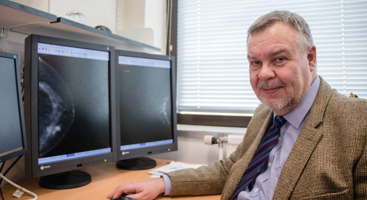
Led by Professor Andrew Evans, Professor of Breast Imaging at the University of Dundee, the researchers tested whether breast cancer cells were still present at surgery and, if so, whether they were detected by US alone, MRI or SWE plus US imaging.
He said, “Our study is the first time that a comparison of this nature has been undertaken and we found that shear wave elastography combined with ultrasound had a similar accuracy to MRI. It’s quick and reproducible so it could potentially replace MRI in the future, which is expensive, time-consuming and cannot be given to some women.
“This is an important finding that could have a real impact on patients’ treatment in the future, and we now need to see larger studies to verify our findings.
“With SWE available on many new machines already in use, if confirmed to be effective in further studies, it may be possible for this method to be adopted quickly across many Scottish cancer centres.”
21 out 80 participants (26%) showed no signs of the tumour remaining when they were assessed at surgery. The researchers found that the combination of SWE and US was as accurate as MRI in identifying whether the chemotherapy had cleared all signs of the tumour in the breast.
With SWE and ultrasound currently being done on the same machine, the authors suggest that the combined imaging technique could also be faster and cheaper than MRI scanning. SWE takes around two minutes to perform and a further two minutes to extract the data. In comparison, the researchers state that an MRI can take between 30 to 45 minutes. Furthermore, a breast MRI is more expensive and is estimated to cost around £250, with US with SWE estimated to cost approximately £50.
The findings, published in journal Clinical Radiology, also suggest that using SWE and US may provide a solution for the 10 per cent of breast cancer patients who are not able to have an MRI scan, such as those who may have a metal object or implant such as a pacemaker.
Dr Kotryna Temcinaite, Research Communications Manager at Breast Cancer Now, which funded the study, said,"We know that having breast cancer surgery can have a significant emotional and physical impact on many patients and there are risks and side effects to surgery. This is a really promising finding that could in future help some patients avoid more extensive breast cancer surgery.
“As more effective treatments become available, this new method could even help some patients to avoid having surgery altogether, which could have a major impact on their quality of life.
“New imaging methods like this will be vital in helping clinicians and patients to decide on the most effective course of treatment, which in turn will help to achieve the best possible quality of life after treatment for breast cancer patients.
“We would encourage anyone who has questions or concerns about breast imaging or breast cancer surgery to speak to their breast care nurse or call our free Helpline on 0808 800 6000.”
For media enquiries contact:
Dominic Glasgow
Media Relations Officer
University of Dundee
Nethergate, Dundee, DD1 4HN
Tel: +44 (0)1382 385131
Email: d.w.glasgow@dundee.ac.uk