13 November 2009
Winning picture shows value of dazzling medical images
Note: Unfortunately Prof Eljamel is not available for interviews until 5.30 pm today at the exhibition opening
The winner in a University of Dundee competition to find stunning pictures arising from biological and medical research shows how advances in imaging are having a dramatic effect in surgery.
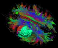
Professor Sam Eljamels three-dimensional image of a malignant brain tumour allowed the successful removal of the tumour through a surgical corridor without affecting the patients speech, motor or sensory functions.
It is now on display in the Dalhousie Building at the University as part of the 'Visions of Discovery' exhibition, which is open to the public from today (Friday 13th Nov).
The image shows the malignant brain tumour as a green ball, surrounded by white matter fibres, with
motor fibres in red, sensory fibres in blue, connecting fibres in green and speech fibres in dark-green behind the tumour.
'What we see in this image is a surgical 'corridor' where we can access the tumour without
damaging any of these vitally important fibres,' said Professor Eljamel, of the Centre for Neuroscience in the Universitys College of Medicine, Dentistry and Nursing.
'That allowed us to carry out the surgery without affecting the patients speech, motor or sensory functions, so it was a very successful surgery.'
Professor Eljamel explained that advances in imaging were leading a new era of personalised treatments for patients.
'It is relatively recently that we have been able to do this kind of thing using such detail,
really over the past year or so,' he explained. 'Previously this kind of surgery depended on a very good knowledge of anatomy, because you had to plan your surgery without having an absolutely clear picture of the best way to reach the tumour without causing any collateral damage.'
'We would have an idea of where the tumour was and where we had to go to get to it. The difficulty of course is that anatomy is not exactly the same for everyone, so it was hard to be absolutely certain of exactly where everything is. An image like this individualises the treatment - you get a clear picture of where everything is for every patient.'
'That means we can have a different surgical plan for each patient, which is a significant improvement.'
Professor Eljamel was named the winner of the Visions of Discovery competition, entries from which form the stunning exhibition. From bacteria to brain tumours, and chromosomes to cancer cells Visions of Discovery shows fascinating images of bioscience research produced by researchers based in the Colleges of Life Sciences and Medicine, Dentistry and Nursing at Dundee.
'Dr Eljamel's winning entry perfectly illustrates how a detailed image can aid understanding of
the underlying medical or scientific issues,' said Dr Jenny Woof, co-organiser of the exhibition and competition, and Reader in Immunology in the Division of Medical Sciences at the University's Medical School, Ninewells Hospital.
'It was a clear favourite among our panel of judges, who scored all the images not only on their aesthetic qualities, but also their originality, informational content, technical proficiency and visual impact. The judges brought a range of perspectives based on their expertise in public engagement, science communication, science publishing and image analysis.'
Judges for the competition were Dr Ken Arnold, Head of Public Programmes, Wellcome Trust; Rose Taylor, Creative Director of the Science Photo Library; Dr. Bernd Pulverer, Editor of Nature Cell Biology; and Professor Anne Anderson, Head of the College of Art, Science and Engineering at the University of Dundee.
Entrants to the competition were encouraged to submit images relating to their research, resulting in a dazzling array of entries reflecting work in subjects ranging from deadly parasites to cells of our immune system, from nerve cells to microtubules in the gut, and from dying cells to fruit fly embryos.
'The subject matter ranges from the large right down to the incredibly small, and from the clinically relevant through to images that illuminate our basic understanding of the cell,' said Dr Paul Andrews, competition and exhibition co-organiser, and a senior scientist in the Drug Discovery Unit in the College of Life Sciences.
'It is worth remembering that while the images are visually quite beautiful and awe-inspiring they are not just pretty pictures - they reflect the cutting edge research taking place in Dundee, using some of the most advanced techniques in the world'
Winners of the competition will be attending a special preview opening of the exhibition from 5.30 pm to 7.00 pm on Thursday November 12th. Prizes will be awarded for the best overall image and the best PhD student entry, among others.
For media enquiries contact:
Roddy Isles
Head, Press Office
University of Dundee
Nethergate, Dundee, DD1 4HN
TEL: 01382 384910
E-MAIL: r.isles@dundee.ac.uk
MOBILE: 07800 581902 |