15 February 2009
Microscope allows Scottish researchers to gain new insights
Researchers in Scotland will be able to explore a microscopic world that until recently has been invisible to the camera or eye, after taking delivery of one of only seven OMX super-resolution microscopes in the world.
The OMX microscope is now in site at the College of Life Sciences at the University of Dundee, where scientists are already using it to gain insight into areas that were previously inaccessible.
More than 400 hundred years have passed since Galileos occhiolino became the first device to be given the name microscope and for the majority of this time the ability of a microscope to distinguish between two points has been limited by the theoretical limit to resolution as defined by Abbe in 1873. The technology used by OMX circumvents this limitation and can generate images with a resolution approximately twice that possible with a conventional light microscope. In real terms, this means that biological structures that were once indistinguishable from one another can now be identified as separate entities.
In designing the OMX, scientists have managed to manipulate light so it is focused into a uniform pattern on the specimen, a method known as structured illumination. Images of multiple cross-sections of the specimen are taken, ultimately revealing previously hidden features of the specimen.
Professor Jason Swedlow, Wellcome Trust Senior Research Fellow at the College of Life Sciences
and one of the main drivers behind getting the OMX to Scotland, explains, 'Its like taking many different cross-section light images of the object under observation and then reconfiguring them to create a detailed full image - a 3D transparency of the object itself, in great resolution.'
The development of light microscopy has been critical in understanding the nature of diseases including cancer and degenerative illnesses. In many cases it has been the imaging of live tissues and cells that has provided the most insight in to our understanding of the biological processes involved. For scientists, this has also provided new challenges when trying to capture images that accurately report on a dynamic process in both space and time.
To meet these challenges OMX has been equipped with very fast cameras and shutters capable of acquiring 100 images per second, a digital signal processor controlling all aspects of image acquisition to ensure precise timing and a stage that can move up and down in steps only 1/10,000th of a mm deep.
The OMX has been brought to Scotland by the Scottish Universities Life Sciences Alliance (SULSA), a strategic partnership between the Universities of Aberdeen, Dundee, Edinburgh, Glasgow, St Andrews and Strathclyde, and the Scottish Funding Council (SFC). By linking researchers together and pooling resources, SULSA provides Scottish life scientists with the state-of-the-art technologies they need to retain their competitive edge.
Professor Mike Tyers, SULSA Director, emphasized the impact of the OMX on Scottish life sciences.
'Through the SFC pooling initiative in the life sciences, and by working together, Scottish universities have acquired a revolutionary technology that few institutes could afford on their own,' said Professor Tyers.
'Scottish researchers now have the opportunity to be the first to visualize many subcellular processes that have never been seen before. The OMX microscope will complement other technology platforms being deployed at other SULSA partners'.
One of the main reasons for locating the OMX at Dundee is the exceptional Microscopy facility
and expertise that already exists here. Dr Sam Swift, Head of Microscopy at the College of
Life Sciences, commented, 'Dundee really is the ideal location for the OMX: we have the facilities, the technology and most- importantly the expertise to let the microscope work to its fullest potential and truly transform Life Sciences Research in Europe.'
NOTES TO EDITORS
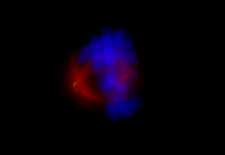
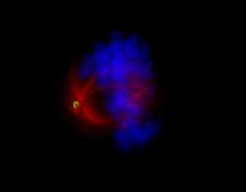
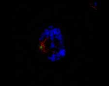
Images - A human cell about to undergo division. Here, the same cell
has been imaged using conventional microscopy, shown without (top)
and with (middle) computational image processing, and using the OMX
microscope structured illumination method (bottom). In the OMX
generated image, it is much easier to see the fine structure of the
microtubules (red) and the pole protein (green) that have important
roles in accurate segregation of DNA (blue) into new cells - a
process that often fails in cancer.
For media enquiries contact:
Roddy Isles
Head, Press Office
University of Dundee
Nethergate
Dundee, DD1 4HN
TEL: 01382 384910
E-MAIL: r.isles@dundee.ac.uk
|