17 October 2005
Microscopy Magic
An enlightening image of a cancer cell has won a researcher at the University of Dundee a top prize in the international Nikon 2005 Small World Photomicrography Competition.
Dr Paul Andrews, of the School of Life Sciences at the University, was one of only two winning entries from the UK, for his submission of an image of cell division.
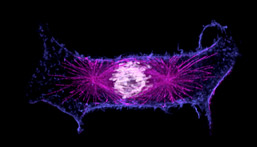
Dr Andrews' prize-winning image shows a cancer cell caught in the act of dividing its chromosomes (shown in white) into two new cells. It does this using the mechanics of the cytoskeleton, which is made up of the microtubule spindle (this part attaches to chromosomes and is shown in purple) and the actin filament network which helps the cell divide into two (shown in blue). Knowing the detailed way in which cells accurately achieve this segregation event is critical for an in depth understanding of cancer.
The International microscopy competition is open to all disciplines of science and received over 1700 entrants. The first twenty prize-winning images are exhibited at numerous museums and science centres throughout the United States.
Dr Andrews is based in the Division of Gene Regulation and Expression in the School of Life Sciences and works on understanding chromosome segregation in cancer cells, using as one of his tools a state-of-the-art digital deconvolution microscope (DeltaVision) for high resolution imaging. /ENDS
Note to Editors:
The Nikon International Small World Competition first began in 1974 as a means to recognize and applaud the efforts of those involved with photography through the light microscope. Since then, Small World has become a leading showcase for photomicrographers from the widest array of scientific disciplines.
For more information contact:
Roddy Isles,
Head of Press
Tel: 01382 344910,
out of hours: 07968298585,
Email: r.isles@dundee.ac.uk |