A unique international exhibition in San Francisco presenting microscope images as art will display six exhibits created at the University of Dundee.
The exhibition, entitled "microModern - an exhibit of art in science, opens tomorrow Thursday 11 December and runs until 20 December at the LIMN Gallery in San Francisco."
Six of the 37 exhibits at the microModern Exhibition are creations of Dr Jason Swedlow and Dr Paul Andrews from the Division of Gene Regulation and Expression and Ms Arwen Wilcock, from the Division of Cell and Developmental Biology in the School of Life Sciences.
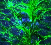
Jason and Arwen's image is of a chick embryo stained for DNA (blue) and microtubules (green). The tissue is from the neural tube, the embryonic structure which develops into the nervous system of the adult animal.
The exhibition of microscopy images is the idea of Paul Millman, Vice-President of Sales for Chroma Technology, who saw the images as art before he saw the science when looking at the stunning images.
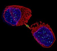
Paul Andrews is exhibiting five images, which show HeLa cancer cells captured in various stages of the cell cycle. The cells were fixed and stained with fluorescent antibodies and also have expressed in them Aurora B protein kinase linked to the jellyfish green fluorescent protein. Paul says "They were taken on a DeltaVision® Restoration Microsocope and then software-enhanced to create an aesthetically pleasing image so some of them bear little resemblance to a real cell!".
Dr Swedlow is a Principal Investigator and Wellcome Trust Senior Research Fellow based in the Wellcome Trust Biocentre where his laboratory is interested in how chromosomes are assembled during cell division. This process is a known target for anti-cancer therapeutics. Jason uses both biochemistry and digital microscopy to understand how chromosomes are organised. In the last few years, they have developed biochemical methods that allow them to directly probe the molecules that are associated with mitotic chromosomes, and have also applied high resolution digital fluorescence microscopy to the study of the structure of the mitotic chromosome.
Dr Andrew's is a postdoctoral scientist in Dr Swedlow's laboratory working on the functions of the Aurora B protein kinase in human cells. Part of these studies involves the use of a state-of-the-art digital deconvolution microscope (DeltaVision®) for high resolution imaging. Ms Wilcock is a Wellcome Trust 4-year Postgraduate Student in the School of Life Sciences. For her postgraduate thesis, she is examining the processes underlying the determination of cell fate during the development of the nervous system. Her project is a collaboration between Dr. Kate G. Storey and Dr. Swedlow. All of this work is funded by the Wellcome Trust.
The exhibition is sponsored by Chroma Technology, who manufacture the specialised optical filters necessary for imaging the multitude of different fluorescent markers used in modern cell biology. Chroma Technology commissioned images from a select group of laboratories from across the globe, all specialising in state-of- the-art imaging. A sample of the exhibition can be viewed online at www.micromodern.com
All proceeds from the sale of images will be donated to the San Francisco AIDS Foundation.
By Jenny Marra, Head of Press 01382 344910, out of hours: 07968298585, j.m.marra@dundee.ac.uk