27 February 2013
Cancer Cell Division conquers GE Imaging Competition
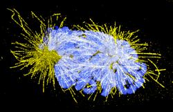
(* Image description: A human cervical carcinoma (HeLa) cell captured in the early stages of cell division. During cell divison the cell needs to ensure the genetic material is equally distributed between daughter cells. Molecular machines working along a dynamic scaffold (microtubules, shown in yellow) separate the genetic material, condensed into chromosomes (blue). This critical process is orchestrated by complex regulatory and control mechanisms, which become defunct in cancer cells.)
A stunning image of a cervical cancer cell in the process of cell division, taken using one of the world's most advanced high resolution microscopes by scientists at Dundee University, has been named the European winner of GE Healthcare's 2012 Cell Imaging Competition.
The image captured by Dr Markus Posch in the College of Life Sciences at Dundee University shows the role that the very latest imaging technology can play in helping scientists to understand the inner workings of human cells.
Dr Posch works with the OMX microscope at Dundee, which uses groundbreaking technology known as 'super resolution microscopy'. This enables structures inside a cell, which play a crucial role in cell division ('microtubules' coloured yellow in the image), to be seen more clearly than ever before, although their diameter is in fact just 1000th of the thickness of a human hair.
"The technology we are now able to use has vastly increased the power of microscopy to the point where we can now see things in the sort of detail that was never before possible," said Dr Posch, who is the Scientific Officer for OMX.
His work is supported by SULSA, the Scottish Universities Life Sciences Alliance, a research pooling partnership between the Universities of Aberdeen, Dundee, Edinburgh, Glasgow, St Andrews and Strathclyde that is supported by the Scottish Funding Council. The OMX has been installed as a national resource and the system has been extremely popular - 50 per cent usage is dedicated to external users, and Dundee has hosted scientists from 19 different institutions from all over the UK and Europe.
"This has tremendous benefits for scientific and medical research as we can view cells in ever greater detail, giving us new understanding of how they work," Dr Posch added.
Now in its sixth year, GE Healthcare's annual competition showcases the beauty of cells and the inspiring research conducted by cellular biologists around the world.
Eric Roman, General Manager of Research and Applied Markets, GE Healthcare Life Sciences, said, "This year's winning images remind us of the cellular complexity behind disease and why the study of cells is so important. We were delighted to receive so many outstanding entries to the competition, which highlights how high-content and super-resolution cell imaging are helping scientists explore the universe of the cell, and so advance our understanding of so many life threatening and life-limiting diseases."
This detailed imaging is playing an important role to aid the discovery of new more effective and targeted drugs to treat diseases such as cancer by stopping diseased cells from replicating.
The technology used is an example of some of the innovations being developed by GE Healthcare which are shaping the face of medicine of the future.
The University of Dundee is one of the first universities in the world to invest in this imaging technology.
It was announced last week that the University of Dundee is to receive around £1million in new funding distributed by three of the UK's research councils to boost the resolution revolution taking place in microscope technology. The money - from the Medical Research Council, the Biotechnology and Biological Sciences Research Council and the Engineering and Physical Sciences Research Council - will support an upgrade to the existing OMX microscope, allowing it to be used for super-resolution imaging in living cells.
For media enquiries contact:
Roddy Isles
Head. Press Office
University of Dundee
Nethergate, Dundee, DD1 4HN
TEL: 01382 384910
E-MAIL: r.isles@dundee.ac.uk
MOBILE: 07800 581902 |