
Visualising the Invisible
A walk along the corridor of Media Arts and Imaging at Duncan of Jordanstone College is a journey to the bottom of the sea, back in time and into the human body.
We are now able to see things that have previously been impossible to view, with Media Arts and Imaging at the centre of a revolution in imaging and animation, developing techniques and methods to see things that there is no other way of seeing.
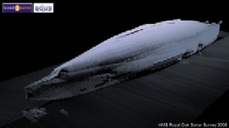
Just under sea level at Scapa Flow in Orkney lies the massive hulk of the Royal Oak, the Dreadnought-class battleships sunk in a U-boat attack in 1939 with the loss of 833 lives. The largest offshore war grave in the northern hemisphere, the wreck continues to leak fuel oil.
The Royal Oak lies in just 30 metres of water, yet no such detailed picture of the wreck has ever been possible to construct until now. New techniques in underwater 3-D visualisation, developed through a partnership between the Universities of Dundee and St Andrews, have produced the first, startling, images of the complete wreck.
"There is just no way you can do something like this using traditional photography," said Chris Rowland, programme leader for animation in the School of Media Arts and Imaging in Duncan of Jordanstone College and a director of the Dundee-St Andrews collaboration ADUS (Archaeological Dive Unit Survey).
"What we have developed is a technique that lets us get a sonar system close to underwater wrecks - or whatever else it is we want to survey - and co-ordinate that with a global satellite positioning system that uses a dozen satellites. The data that is produced lets us build these 3-D visuals with great detail."
The ADUS images of the Royal Oak clearly show the impact damage of the four torpedoes that sank the ship. One of the most powerful images is of the starboard side of the upturned hull in which a bite appears to have been taken out of the bow where the first torpedo struck shortly before 1am as the crew slept. At the other end of the 620ft hull the rudder mechanism is serenely unaffected.
The ADUS team were commissioned by the Ministry of Defence to carry out the sonar survey of the wreck. "The MoD were keen to see what sort of condition the hull was in after 67 years on the seabed and whether there was a danger of it cracking up," said Chris.
"To do that through a series of dives would take a lot of time and you still wouldn't end up with as clear and complete a picture as we can produce using the sonar system. In the right conditions we can produce a picture like this in one day."
"These are images which are produced in 3-D, and we can use these to do a virtual `flypast' and examine any features. This leads to a much better understanding of the state of the wreck than can be gleaned from static print images."
The system is being continually refined and updated by the team - "we are absolutely research-driven" said Chris - "but the potential is already obvious. The team have already been commissioned to carry out surveys of other wrecks and there are potential uses for everything from the oil industry to recreational diving."
Uncovering what lies underwater is only one of the areas where the techniques used in animation and imaging are helping break new ground.
It is now not unusual to find staff from Life Sciences, Medicine or Engineering striding the corridors of the art college as link-ups form to develop new applications which are opening up areas which previously were hidden from our view.
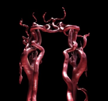
Two of the key threads being pursued at present are in medical imaging and forensic art. In medical imaging, significant advances are being made in developing images that can be shown both to clinicians and their patients. The quality of images being developed is anticipated to greatly aid communication between doctor and patient.
"Our post-graduate students like John McGhee and Emma Fife have been working hand-in-hand with clinicians to develop these new systems and applications which offer real potential in breaking new ground in showing how things work inside the body," said Chris.
"It is very much a collaborative effort - the emphasis is very much on both ends of this contributing to make something new. It couldn't work without the involvement of both the artists and the specialists in their own fields, in this case medicine."
John McGhee has been working closely with clinicians and nurses in NHS Tayside, particularly within the Radiology department at Ninewells Hospital, on developing 3-D images of the body's innermost workings.
The images produced are stunning. Blood cells are pumped through veins, kidneys are represented in vivid colour, and the sculpture of our insides is brought to life, in pictures which are finding a home in the rooms of hospital consultants and art galleries.
"There is a broad audience for this work, from the medical community and the practical applications there, to a general public who are interested in the body and how it works, and on to the artistic community," said John.
"The focus has been on developing something which is of real use in the clinical setting. It is designed for use in one-to-one patient consultation and we have interviewed around 20 patients at Ninewells, with really positive results."
"The response from the clinicians and nurses we have spoken to at NHS Tayside - who have been immensely supportive of my work - has been that this is something which could be used to help change people's behaviour in relation to their own health. If we were to achieve that, then that would be a major step forward."
"But there is also a wider value to the work. We are using aesthetics to help people understand the body. People do have a real appetite for understanding the body and its structure, we can see that just from the amount of television programmes which cover that kind of ground."
"What we do is place the information about what happens within the body into the sort of technology more familiar to Hollywood or computer games, and it gives a very strong image which is clearly understandable. The kind of 2-D scans used in hospitals just now are not particularly clear to people without clinical expertise. However, using them in conjunction with the 3-D images I have produced builds a much clearer picture."
Both John's images of the body and the underwater images of the Royal Oak are examples of the new fields of vision that are being opened up for us. The invisible is being given form and, more importantly, function.
Next Page
Return to December 2006 Contact
