
Remote controlled medicine
Ultrasound and lasers could give surgery the chop
Going under the knife for medical treatment, such as the removal of a tumour, could one day be a thing of the past as the surgeon's scalpel is replaced by ultrasound and lasers in an exciting new area of research.
'Sonoptics' is an emerging branch of science that explores the possibility of using light and sound waves outside the body to manipulate cells inside the body.
The most exciting potential for the technology is in the area of medicine, where lasers and ultrasound have been shown to have the capability of opening a cell's membrane so that it allows therapeutic agents such as medicines to flow into the cell, whereas previously the membrane would have remained closed.
A research team based at the University of Dundee and St Andrews has already made some major advances in understanding how lasers and ultrasound can influence disruption to cell membranes and are now embarking on a major project to develop novel techniques which they hope could one day see the end of traumatic, invasive surgery.
The past number of years has seen some major advances in the development of specific molecular therapeutics such as genes and drugs. The idea being that these species will then travel to the site in the body where they are needed and can then be somehow absorbed by the damaged or infected cell so as to elicit their therapeutic action.
However, there are some stumbling blocks to this approach. The first is how to ensure that the molecular therapeutic agent is taken up only by the infected or dysfunctional cell and not other healthy cells around it. For example, if the aim is destroy a cancerous tumour in the liver, it is not desirable for the healthy tissue surrounding the tumour to also be destroyed.
Secondly, human cells are naturally designed not to allow foreign molecules in except under very specific membrane barrier control.
Both ultrasound and lasers have been shown to overcome these problems.
Ultrasound and microbubbles
Dr Paul Campbell in the University of Dundee's School of Engineering and Physical Sciences is leading the research into the ultrasound side of the project, which has already resulted in a major publication in Nature Physics.
"It had been known for some time that cells exposed to extremely high-frequency sound waves, or ultrasound, become more permeable and capable of taking up large molecules, such as genes or drugs."
"However, how this happened was not understood and without this understanding it was extremely difficult to move forward and develop any potential technologies for treatment," Dr Campbell said.
"Our research was the first to clearly illustrate what happens when ultrasound and microbubbles interact and how they impact on a cell's membrane to make it more porous."
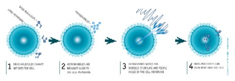
Microbubbles are tiny, gas-filled bubbles that are about half the diameter of human red blood cells and thus small enough to pass through even the finest capillaries of the blood's circulatory pathway.
Scientists often use microbubbles in conjunction with ultrasound therapy because they have the effect of amplifying the sound waves reflection property. Effectively, the bubbles create a stronger echo when the ultrasound wave passes through them and in this way the signal is enhanced considerably. This improves contrast in regular ultrasound imaging and allows easier recognition of certain tissues and organs.
Dr Campbell and his team used laser light in a technique called "optical trapping" to control the location of the microbubbles, moving them close to a specific target cell. They showed that when ultrasound was passed through them, the microbubbles first inflate slowly then rapidly collapse, forming thin, needle-like projections which then pierced the near-most cell membrane, opening a door to the cell and making it porous.
At high intensities of ultrasound they were able to kill the target cell; with lower intensities they could simply make the cell more permeable.
"Our findings open up the potential for two different uses for this technology. Firstly, it could be used to remotely kill infected or cancerous cells. Another potential use is to make cells more permeable and therefore more capable of taking up drugs or other therapeutic molecules," Dr Campbell said.
Blue laser
Led by Professor Kishan Dholakia, the St Andrews-based team have already successfully shown the ability to target and perforate specific single cells without effecting surrounding cells. Their method has also resulted in DNA being taken up by the laser-perforated cell, proving the potentials of the technology for therapeutics.
Their technique is believed to be the simplest and most powerful laser method developed to date.
"This dual approach technology allows us, in principle, to inject any molecule into any cell. Indeed, we have shown that even genetic material can be introduced into cells using the laser-based approach with successful downstream biological effects," said Professor Dholakia.
The team now plan to further develop their methodology, test the technique on different cell types and investigate further what is happening at the cellular level when the laser acts on the cell membrane.
Laser light and ultrasound: where to now?
Both ultrasound and lasers have been shown to have the ability to permeate, or even kill, a cell in a very controllable and reliable way. But the two techniques are very different.
"At this stage, there are merely suggestions as to which method will turn out to be the more successful. It may be that both are useful but in different situations," Dr Campbell said.
"For example, at high intensities, ultrasound has the potential to cut a cell-wide channel through a tumour, opening it up so that it is weakened from the inside. On the other hand, lasers can be more gentle and could be used to specifically target certain cells. Of course, the two may also complement each other and there may be times when both can be used."
The research team was recently awarded £2 million through the UK 'Basic Technology' Programme, administered by the Engineering and Physical Sciences Research Council, to further develop their research.
Their first plan is to fine tune their ultrasound and laser techniques to find the best parameters for allowing specific therapeutic agents or genes into specific cell types. They will then go on to investigate and develop specific studies for the treatment of diseases such as cancer and neurobiological disorders.
Dr Campbell said, "The overriding objective for this project is to revolutionise the activation and delivery of genes, drugs and therapeutic molecules into live biological materials."
"Developing a means to controllably deliver drugs at remote anatomical sites, yet in a very non-invasive way, is a significant challenge of heightened academic and industrial interest. This is underscored by the market for delivery technologies which is estimated to be around 30 billion dollars in the US."
"Sonoptics is likely to become a major area of research over the next decade and one we believe will lead to a quantum leap for medical research."
Next Page
Return to October 2006 Contact
